
Biometric Method for the
Ossification Evaluation of Children from Birth Up to the Ages of Two and Four –
Applied to the Metacarpal and Phalanxes in Spanish Longitudinal Series
Original
Article published 06/26/2013
Bernardo
Ebri1 , Inmaculada Ebri1
Affiliations
- 1. Hospital Universitario
Miguel Servet de Zaragoza (Spain)
DOI
Cite as
Ebri B, Ebri I (2013) Biometric Method for the Ossification Evaluation of
Children from Birth Up to the Ages of Two and Four – Applied to the Metacarpal
and Phalanxes in Spanish Longitudinal Series. Cureus 5(12): e151.
doi:10.7759/cureus.151
Disclosure/COI
The authors have no conflict of interest to disclose.
Copyright
© 2013 Ebri et al.
License
This is an open access article distributed under the terms of the Creative
Commons Attribution License, which permits unrestricted use, distribution, and
reproduction in any medium, provided the original author and source are
credited.
Aim: This work, based on the
Spanish longitudinal growth and development series done at the Andrea Prader
Center, Zaragoza, Spain, studied children up to the age of twenty years. It
aims to contribute to a practical and accurate numerical method to calculate
the bone age of the studied children, from birth to two and four years.
Methods: The total sample of
the study was 160 healthy children (73 males and 87 females). Every child
underwent annual radiography on his/her left hand at the Miguel Servet Hospital
in Zaragoza, Spain. Using measurements of the Tanner II-Rus method, the maximum
epiphyseal distances of the subjects were studied.
Results: As a result, we have
developed an index called the Metacarpal-Phalanx Index, closely correlated with
the chronological age of the child, which creates bone age prediction
equations. Another index called “Index Valuation Ossification of the
Metacarpal-Phalangeal", obtained through the above, allowed us to compare
the results to Gaussian shape equivalences, thus revealing the ossifying status
of the child, and whether it is late, early or insignificant.
Conclusions: When using this
method, we are able to optimize the calculations of bone age if we apply the
general equations to children up to the age of twenty years.
Introduction
This work's main objective is
to provide a numerical method to accurately calculate the bone age of children
in the age groups from birth to two and to four years of age. This study is
based on the Spanish longitudinal growth and development series, "Andrea
Prader" [1], which assesses children from birth to twenty years. The
numerical method evaluates the epiphysis of the metacarpal bones and phalanges
of the left hand in the same bones of Tanner, et al. [2] but rather by
following another dynamic assessment. In our method, we measure the maximum
distance from the nuclei of ossification and prepare them as average indices
(Ebrí índices), designated as the "Indice Metacarpo-Falangico" (IMF)
or “Metacarpal Phalangeal Index” and the "Ossification Valuation Index of
the Metacarpal-Phalangeal" ( IVOMF )
Ebrí Torne [3] published the
"Ossification Evaluation Index of the Tarsus" (OEIT) applied to a
Spanish cross-population from birth to age 16, and later, also published [4]
the same rate applied to children up to two and four years. The latter
publication was better at this age for the predictive equations of general
casuistry, relativizing the asynchrony of the nuclei of ossification, and
avoiding overestimation of bone age that produced general equations when
applied to children. Similarly, in this present work with the
metacarpal-phalangeal region, we wanted to provide the practical predictive
equations for optimizing bone age prediction for these age groups.
Materials
& Methods
The total sample of the study
were 160 healthy children (73 males and 87 females) whose left hand was
radiographed annually on his or her birthday, at the Hospital Miguel Servet in
Zaragoza (Spain), from birth to four years, inclusive. The Research Committee
authorized this study by the General Study and Radiological Somatometric Andrea
Prader of the Unit of Endocrinology, Hospital Miguel Servet, Zaragoza, Spain.
Signed consent was obtained from the parents of all the children. The study is
also endorsed and supported by the Government of Aragon [5].
Action films: From birth to
two years, there were 162 males and 201 females. From birth to four years,
there were 255 males and 326 females. We used the 1956 survey Graffar
socioeconomic classification for children [6].
The procedure in every left
hand radiograph was to measure the core of the metacarpal and phalangeal
epiphyses comprising both the radius and ulna. Maximum distances were measured
with vernier nuclear optimally expressing the measurements made and have served
as the basis for preparation of the IMF. Such index is expressed in millimeters
(mm), and its result, the sum of the maximum diameters of the cores epiphyseal
metacarpals and phalanges: I, III and V, as well as the radial and ulnar distal
epiphysis in a total 13 cores.
Figure 1 shows the maximum
distance measured from the cores of ossification.
Figure 1:
Maximum
diameters of the Metacarpal ossifying cores
The sum of the existing cores
at the time of radiographic measurement is divided by 13, for simplification of
the index number. A fixed number, in all cases, even at the time of
measurement, was not present in all cores. All radiographic measurements were
performed by a single observer. A repeatability study performed in 100 films,
one month after the first measurement, was greater than 95%. The statistical
package, "Statistix software version 9, 2000", was used for
statistical work.
Correlation coefficients were
made of the two variables, IMF and chronological age, in order to obtain the
equation of the regression line for the prediction of the child's bone age. For
better optimization of reading the bone age of the child to study, and Gaussian
bell graphic expression, was obtained from IMF index, called the
"IVOMF" (Ossification Valuation Index of the Metacarpal-Phalangeal),
following the same general methodology that Torne Ebrí used in various studies
[7-8]. This is the formula applied by this author:
a + 1, 96 sd + b.Index - Age
IVO =
-------------------------------------
0, 0392 sd
Results
Most of the study population belongs
to socioeconomic status III (middle-middle class), 46.2%. Category IV (lower
middle class) represents 43.9%.
The equations of straight bone
ages for male and female, of newborn to two years and newborn to four years,
are expressed in Table 1. Table 1 also specifies the correlation coefficient,
number of radiographs, and standard deviation. The statistical significance of
the correlations is: p <0 .001.="" o:p="">
Table 1:
Table 1. Equations to find out the Bone Age.
Bone Age = a + b. IMF
Table 2 specifies the IVOS
formulas for children.
Table 2:
Table 2. Equations to obtain the MF-Ebrí ossifying valuation index
0-2 años. Male. IVO-IMF= 100,919 +26,796 x IMF -61,711 x EC
0-4 años. Male. IVO-IMF = 93,049 +26,259 x IMF -48,978 x EC
0-2 años. Female. IVO-IMF = 112,492 +31,419 x IMF -96,167 x
EC
0-4 años. Female.
IVO-IMF = 85,244 +29,554 x IMF -62,294 x EC
EC = Chronological
Age in years.
IMF = Metacarpal-phalanx index in mm,
obtained from the radiological measurements.
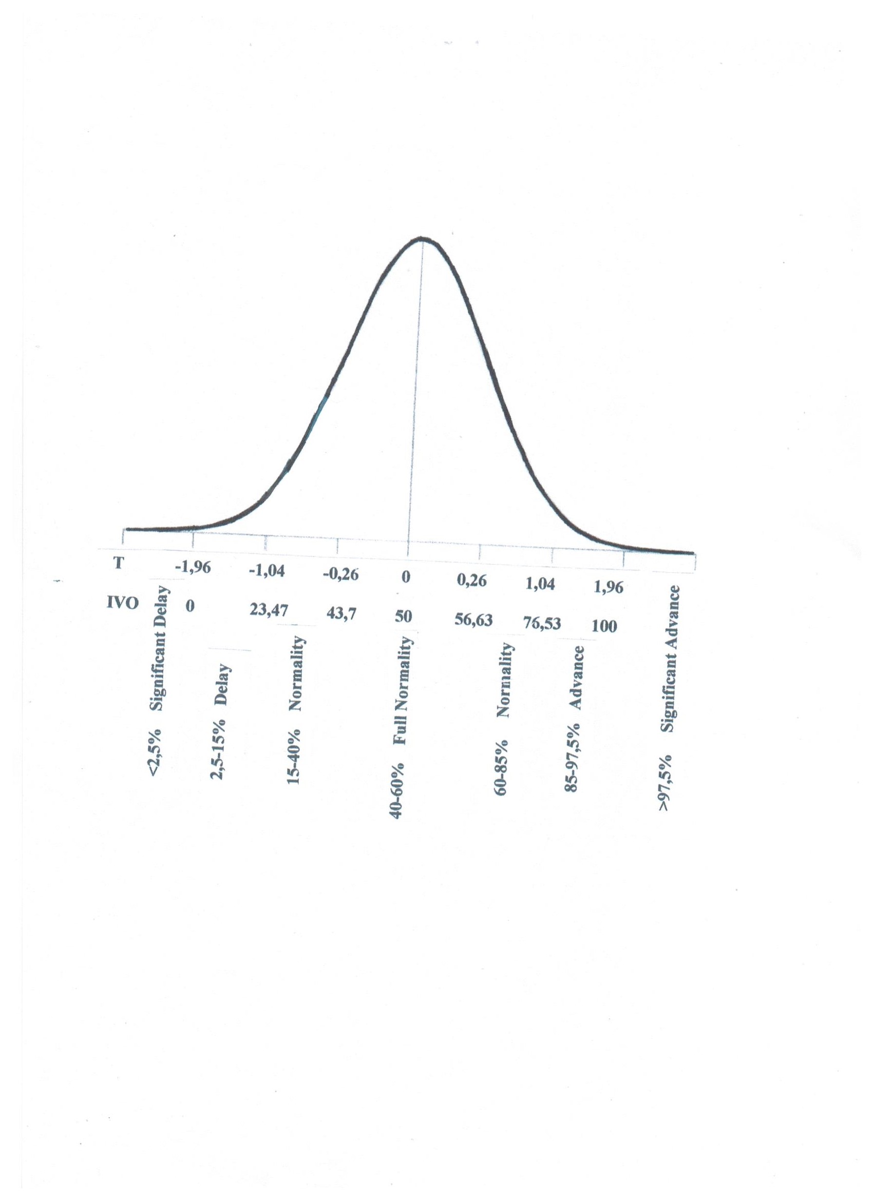
The results obtained, the
values in both sexes, ranging from 0 to 100 (-1.96 to 1.96 standard
deviation) can be brought to a figure of equivalence regarding bone age (Figure
2). The radiograph would show, in this way, a normal, advanced, or delayed
ossification and if the lead or lag is significant or not.
Figure 2:
IVO
equivalences to the bone age
Figure 2 specifies the IVO
equivalences to the bone age.
Discussion
Bone age assessment is
frequently used in endocrine pathology to assess milestones of nutrition and
growth, as well as to serve as a modern method of predicting adult height and
to check the response to suitable treatment for pathologies that may accelerate
or retard normal growth. Accurate bone age can also be applied in anthropology,
forensics, and school sports, as well as to control children adopted by
institutions [9]. IVOMF and IMF are accurate means for bone age determination,
allowing the investigator two effective tools for diagnosing predictive bone
age in study children. By adjusting the IVOMF at ages newborn to two years and
newborn to four years, excluding the rest of older casuistry, the asynchrony of
the nuclei of ossification is relativized. This avoids the overestimation in
the usual general equations that has occurred when applied to young children.
With the figure of equivalences IVO bone age, the diagnosis of bone age can
further be simplified. A simple pocket calculator provides a substantial
improvement for bone age assessment, so important in the overall assessment of
the child.
Ebrí developed the study's
methodology in 1992 and 1993 in the application of comparing a longitudinal
Swiss population to a Spanish population by using the methods of Greulich and
Pyle with those of Tanner-Whitehouse [10-11]. In 1996, Ebrí [12] applied his
Swiss longitudinal population indices, the same bone Rus TWII studied, by
comparing the two methods, checking their compatibility, and found a greater
simplicity of bone assessment index metacarpofalángico compared to complex
methodology of the English author. A year later Ebrí [13], in the same Swiss
population of 10-22 years, made a comparative study of bone ages of these
children by different methods: carpal Tanner, Tanner-Rus, Ebrí carpal, Ebrí
metacarpofalángico, and Greulich-Pyle . He checked the concordance between
them, even detecting differences, since each methodologically behaved
differently. Preference was chosen - the same bones that TWII-Rus analyzed were
preferred, as they were more useful than the carpals and correlated better with
pubertal changes, being most predictive of adult height [14].
Also, the basic methodically
calculated bone age, as presented here, can also be applied prospectively in
order to study new or different racial groups for the purpose of creating
standards.
Conclusions
The author presents an
accurate and yet simple method to obtain children's bone age in this age range,
avoiding the difficulties introduced by asynchronies occurring in only one age.
1. Ebrí Torné B, Ebrí Verde B: Ossifying Value Index: Ebrí Metacarpal-phalanx in
Aragon lengthwise series (Andrea Prader). An Pediatr (Barc) 2011, doi: 10.1016/j.anped.2011.01.032
2. Tanner JM, Whitehouse RM, Marshall WA, Healy MJ, Goldstein H: Assessment of skeletal maturity and prediction of
adult height (TW2 method) London: Academic Press; 1975.
4. Ebrí Torné B: Biometric method for the ossification evaluation of
children from birth up to the ages of two and four-applied to the tarsus. Acta
Paediatr 1993, 82:872.
5. Ferrández Longás A: Lengthwise Study of Normal Spanish Children from Birth
to Adult Age: Anthropometric, pubertal, radiologic, and intellectual data. Zaragoza:
Andrea Prader Foundation; 2005.
7. Ebrí Torné B: Bone maturation on carpal and tarsus. Radiological
infant clinical study on 5225 children. Zaragoza:
Heraldo de Aragón Publishing; 1988.
8. Ebrí Torné B., Altarriba Farrán J: Presentation of a new biometric method (I.V.O.) for
bone age valuation in children. Medizinische Klink 1979, 214:50-56.
10. Ebrí Torné B: Comparative study of Greulich & Pyle American
Atlas of Swiss and Spanish population, through the Bone Age calculation methods
of Greulich & Pyle, and Ebrí carpal. Annals of Miguel Servet Hospital
1992, 4:83-90.
11. Ebrí Torné B: IVO Carpal of Swiss Child (Lengthwise Study of Zurich). Acta Pediatr
Esp 1993, 51:651-654.
12. Ebrí Torné B: Metacarpal-phalanx bone valuation index. Swiss
Lengthwise Study. Comparative
study with Tanner II Rus. Acta Pediatr Esp 1996, 54:94-102.
13. Ebrí Torné B: Comparative study of bone ages Tanner-Rus, Tanner
Carpal, Ebrí carpal, Ebrí Metacarpal-phalanx, and Greulich & Pyle. Acta Pediatr
Esp 1997, 55:369-374.
14. Ebrí Torné B, Ebrí Verde I: Metacarpal-phalanx and carpal numerical index for bone
age calculation and adult height prediction. An Pediatr (Barc) 2012, 76:199-213.
Ebri B, Ebrí I (2013)
Biometric Method for the Ossification Evaluation of Children from Birth Up to
the Ages of Two and Four – Applied to the Metacarpal and Phalanxes in Spanish
Longitudinal Series. Cureus 5(12): e151. doi:10.7759/cureus.151


No comments:
Post a Comment
Añadir comentarios al blog, si queréis aportar alguna opinión